
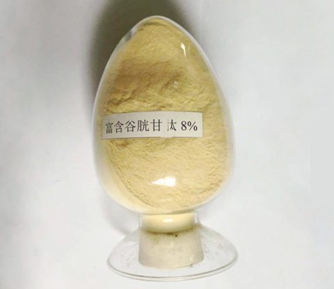
L-glutathione Enriched Yeast Extract


【Other names】: L-Glutathione Reduced, GSH
【Specification】: 8%, 15%, 50%, 98% HPLC
【CAS No. 】: 70-18-8
【Molecular formula】: C10H17N3O6S
【Molecular weight】:307.33
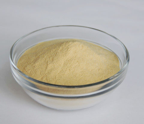
【Appearance】: Yellowish to yellow-brown powder(8%, 15%); Offwhite(50%),White powder(98%)
【Particle size】: 95% pass 80 mesh size

Brief Introduction
Glutathione (GSH) is an important antioxidant in plants, animals, fungi, and some bacteria and archaea. Glutathione is capable of preventing damage to important cellular components caused by reactive oxygen species such as free radicals, peroxides, lipid peroxides, and heavy metals. It is a tripeptide with a gamma peptide linkage between the carboxyl group of the glutamate side chain and the amine group of cysteine, and the carboxyl group of cysteine is attached by normal peptide linkage to a glycine.
Thiol groups are reducing agents, existing at a concentration around 5 mM in animal cells. Glutathione reduces disulfide bonds formed within cytoplasmic proteins to cysteines by serving as an electron donor. In the process, glutathione is converted to its oxidized form, glutathione disulfide (GSSG), also called L-(–)-glutathione.
Once oxidized, glutathione can be reduced back by glutathione reductase, using NADPH as an electron donor. The ratio of reduced glutathione to oxidized glutathione within cells is often used as a measure of cellular oxidative stress.
Difference between Glutathione Reduced(GSH) and Oxidized(GSSG)
Glutathione is a protein made from three amino acids: glutamate, cysteine and glycine. It is a powerful antioxidant formed in the liver, and it supports a wide variety of immune functions throughout the body. Glutathione can exist as one of two different forms: glutathione and L-glutathione.
L-glutathione also is known as reduced glutathione, or GSH, and is composed of a single glutathione molecule with the addition of a sulfhydryl group. When the sulfhydryl groups of two reduced glutathione molecules become oxidized through the loss of electrons, the two molecules can become linked by a disulphide bond and form oxidized glutathione, or GSSG. Oxidized glutathione then is cycled back into reduced glutathione by the glutathione reductase enzyme.
Reduced glutathione is the active form. It donates electrons to free radicals, or molecules with unpaired electrons. The addition of donated electrons neutralizes the free radicals and prevents them from causing cellular damage. Glutathione also participates in various other cellular processes, such as acting as a co-enzyme in biological reactions.
Benefits of Glutathione
Glutathione exists in both reduced (GSH) and oxidized (GSSG) states. In the reduced state, the thiol group of cysteine is able to donate a reducing equivalent (H++ e−) to other molecules, such as reactive oxygen species to neutralize them, or to protein cysteines to maintain their reduced forms. With donating an electron, glutathione itself becomes reactive and readily reacts with another reactive glutathione to form glutathione disulfide (GSSG). Such a reaction is probable due to the relatively high concentration of glutathione in cells (up to 7 mM in the liver).[1]
Generally, interactions between GSH and other molecules with higher relative electrophilicity deplete GSH levels within the cell. An exception to this case involves the sensitivity of GSH to the electrophilic compound’s relative concentration. In high concentrations, the organic molecule Diethyl maleate fully depleted GSH levels in cells. However, in low concentrations, a minor decrease in cellular GSH levels was followed by a two-fold increase.[2][3]
GSH can be regenerated from GSSG by the enzyme glutathione reductase (GSR):[3] NADPH reduces FAD present in GSR to produce a transient FADH-anion. This anion then quickly breaks a disulfide bond (Cys58 – Cys63) and leads to Cys63’s nucleophilically attacking the nearest sulfide unit in the GSSG molecule (promoted by His467), which creates a mixed disulfide bond (GS-Cys58) and a GS-anion. His467 of GSR then protonates the GS-anion to form the first GSH. Next, Cys63 nucleophilically attacks the sulfide of Cys58, releasing a GS-anion, which, in turn, picks up a solvent proton and is released from the enzyme, thereby creating the second GSH. So, for every GSSG and NADPH, two reduced GSH molecules are gained, which can again act as antioxidants scavenging reactive oxygen species in the cell.
In healthy cells and tissue, more than 90% of the total glutathione pool is in the reduced form (GSH) and less than 10% exists in the disulfide form (GSSG). An increased GSSG-to-GSH ratio is considered indicative of oxidative stress.[4]
Glutathione has multiple functions
1). It maintains levels of reduced glutaredoxin and glutathione peroxidase[5]
2). It is one of the major endogenous antioxidants produced by the cells, participating directly in the neutralization of free radicals and reactive oxygen compounds, as well as maintaining exogenous antioxidants such as vitamins C and E in their reduced (active) forms.[6][7][8]
3). Regulation of the nitric oxide cycle is critical for life, but can be problematic if unregulated.[9]
4). It is used in metabolic and biochemical reactions such as DNA synthesis and repair, protein synthesis, prostaglandin synthesis, amino acid transport, and enzyme activation. Thus, every system in the body can be affected by the state of the glutathione system, especially the immune system, the nervous system, the gastrointestinal system, and the lungs.
5). It has a vital function in iron metabolism. Yeast cells depleted of GSH or containing toxic levels of GSH show an intense iron starvation-like response and impairment of the activity of extramitochondrial ISC enzymes thus inhibiting oxidative endoplasmic reticulum folding, followed by death.[10]
6). It has roles in progression of the cell cycle, including cell death. GSH levels regulate redox changes to nuclear proteins necessary for the initiation of cell differentiation. Differences in GSH levels also determine the expressed mode of cell death, being either apoptosis or cell necrosis. Manageably low levels result in the systematic breakage of the cell whereas excessively low levels result in rapid cell death.[11]
Reference:
1. Kaplowitz, N. (1981-01-01). “The importance and regulation of hepatic glutathione.”. The Yale Journal of Biology and Medicine. 54 (6): 497–502. ISSN 0044-0086. PMC 2596047 . PMID 7342494.
2. Jump up^ Bannai, Shiro; Tateishi, Noriko (1986-02-01). “Role of membrane transport in metabolism and function of glutathione in mammals”. The Journal of Membrane Biology. 89 (1): 1–8. doi:10.1007/BF01870891.ISSN 0022-2631.
3. Jump up^ Bannai, S. (1984-02-25). “Induction of cystine and glutamate transport activity in human fibroblasts by diethyl maleate and other electrophilic agents”. The Journal of Biological Chemistry. 259 (4): 2435–2440.ISSN 0021-9258. PMID 6142042.
4. Jump up^ Halprin, Kenneth (1967). “The Measurement of Glutathione in Human Epidermis using Glutathione Reductase”. Journal of Investigative Dermatology. 48 (2): 149. doi:10.1038/jid.1967.24.
5. Jump up^ Grant, Chris M. (2001-02-01). “Role of the glutathione/glutaredoxin and thioredoxin systems in yeast growth and response to stress conditions”.Molecular Microbiology. 39 (3): 533–541. doi:10.1046/j.1365-2958.2001.02283.x. ISSN 1365-2958.
6. Jump up^ Dringen, R. (2000-12-01). “Metabolism and functions of glutathione in brain”. Progress in Neurobiology. 62 (6): 649–671. doi:10.1016/s0301-0082(99)00060-x. ISSN 0301-0082. PMID 10880854.
7. Jump up^ Scholz, RW. Graham KS. Gumpricht E. Reddy CC. (1989). “Mechanism of interaction of vitamin E and glutathione in the protection against membrane lipid peroxidation”. Ann NY Acad Sci. 570: 514–7.doi:10.1111/j.1749-6632.1989.tb14973.x.
8. Jump up^ Hughes, RE (1964). “Reduction of dehydroascorbic acid by animal tissues”. Nature. 203 (4949): 1068–9. doi:10.1038/2031068a0.
9. Jump up^ Clementi, Emilio; Smith, Guy Charles; Howden, Martin; Dietrich, Salvador; Bugg, S; O’Connell, MJ; Goldsbrough, PB; Cobbett, CS (1999).”Phytochelatin synthase genes from Arabidopsis and the yeastSchizosaccharomyces pombe”. The Plant cell. 11 (6): 1153–64.doi:10.1105/tpc.11.6.1153. JSTOR 3870806. PMC 144235 .PMID 10368185.
10. Jump up^ Kumar, Chitranshu; et al. (2011). “Glutathione revisited: a vital function in iron metabolism and ancillary role in thiol-redox control”.The EMBO Journal. 30: 2044–2056. doi:10.1038/emboj.2011.105.PMC 3098478 . PMID 21478822.
11. Jump up^ Hall, A. G. (1999-03-01). “Review: The role of glutathione in the regulation of apoptosis”. European Journal of Clinical Investigation. 29(3): 238–245. doi:10.1046/j.1365-2362.1999.00447.x. ISSN 0014-2972.PMID 10202381.